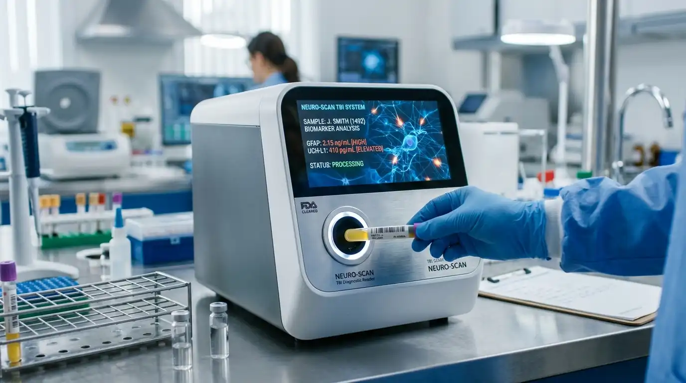
Among the most challenging personal injury cases in California are those involving “invisible” brain injuries—specifically mild Traumatic Brain Injuries (mTBIs), concussions, and diffuse axonal injuries. Insurance defense adjusters routinely exploit standard hospital imaging, pointing to normal CT scans and conventional MRIs to argue that an crash victim is uninjured or exaggerating their symptoms. Today, the intersection of advanced forensic neuroimaging and biomechanical reconstruction is transforming how traumatic brain injuries are proven in court.
1. The Limitations of Conventional Imaging in Mild TBI Cases
 In the immediate aftermath of a motor vehicle collision or severe slip-and-fall, emergency room physicians rely primarily on Computed Tomography (CT) scans and standard 1.5T Magnetic Resonance Imaging (MRI). These structural diagnostic tools are designed for acute triaging—specifically detecting life-threatening skull fractures, intracranial hemorrhages, epidural hematomas, and massive tissue displacement.
However, mild traumatic brain injuries rarely involve gross structural bleeding or focal brain lesions. Instead, the rapid acceleration, deceleration, and rotational forces of an impact cause microscopic shearing and stretching of delicate nerve fibers throughout the brain’s subcortical tissue—a condition known as diffuse axonal injury (DAI).
Because microscopic axonal shearing occurs at a cellular level, standard structural CT and MRI scans pass straight through damaged tissue, returning “unremarkable” or “normal” diagnostic results. Defense attorneys frequently weaponize these normal emergency room scans to label cognitive deficits, memory loss, executive dysfunction, and emotional changes as non-existent or psychological.
2. Diffusion Tensor Imaging (DTI): Visualizing White Matter Disruption
To bring objective evidence of cellular damage before a jury, plaintiff attorneys utilize advanced neuroimaging techniques, chief among them Diffusion Tensor Imaging (DTI).
DTI is an advanced magnetic resonance technique that maps the microstructural integrity of the brain’s white matter tracts by measuring the directional movement of water molecules (anisotropy). In healthy, intact neural pathways, water molecules move predictably along the longitudinal axis of nerve fibers (high fractional anisotropy). When shear forces tear or damage the structural myelin sheath of these axonal tracts, water molecules diffuse abnormally outward (fractional anisotropy decreases).
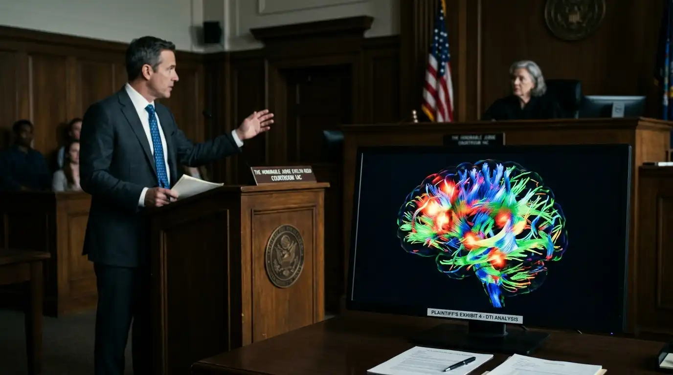
By processing these water diffusion metrics through specialized software, neuroradiologists generate 3D color-coded fiber tractography. DTI transforms subjective patient complaints—such as brain fog, delayed processing speed, and severe headaches—into clear, color-graphic proof of physical neural tract damage.
According to extensive clinical research indexed in the National Center for Biotechnology Information DTI TBI Study, advanced DTI metrics demonstrate exceptional sensitivity in identifying traumatic white matter disruption that standard structural MRIs miss completely.
3. Biomechanical Reconstruction: Connecting Impact Forces to Tissue Damage
While DTI scans establish the physical presence of neural pathway damage, defense experts often attempt to attribute these abnormal findings to pre-existing conditions, aging, or unrelated medical history. This is where biomechanical accident reconstruction becomes indispensable.
Biomechanical engineers analyze physical crash evidence, event data recorder (EDR) “black box” readings, vehicle damage profiles, and occupant dynamics to calculate the exact physical forces applied to the victim’s head during the collision. Key biomechanical metrics include:
- Linear and Angular Acceleration: Calculating the exact G-forces exerted on the skull during impact.
- Rotational Velocity and Shear Strain: Demonstrating how rapid head rotation causes internal brain tissue to slide across rigid internal cranial ridges.
- Head Injury Criterion (HIC): Correlating physical impact severity with established medical thresholds for traumatic axonal deformation.
By combining 3D computer simulations with physical injury thresholds, biomechanical experts establish a direct causal bridge showing that the specific physical forces generated in the crash were more than sufficient to cause the microstructural brain damage revealed on the plaintiff’s DTI scans.
4. Admissibility in California Courts: The Sargon and Kelly-Frye Standards
Introducing cutting-edge forensic evidence in California state courts requires navigating strict evidentiary gatekeeping rules. Defense counsel routinely file pre-trial motions in limine to exclude DTI scans, claiming the technology is experimental or unproven for clinical diagnosis.
Under California law, expert opinion testimony must comply with California Evidence Code Section 801 guidelines, which dictate that expert opinions must be grounded in reliable matter and sound methodology. Furthermore, trial judges act as strict gatekeepers pursuant to the landmark California Supreme Court decision in Sargon Enterprises, Inc. v. University of Southern California (2012).
To satisfy the Sargon gatekeeping standard and the Kelly-Frye rule for scientific reliability:
- General Acceptance: Experienced trial attorneys establish that DTI technology is FDA-cleared and widely accepted across major clinical research institutions (such as UCSF, UCLA, and Harvard Medical School).
- Qualified Experts: The testimony must be delivered by credentialed board-certified neuroradiologists, neurologists, and forensic biomechanical engineers who utilize standardized normative database comparisons.
- Case-Specific Foundation: Experts must connect scan findings directly to the patient’s clinical history, formal neuropsychological testing results, and physical collision dynamics.
5. Overcoming Defense Tactics and Securing Fair Recovery
When faced with objective DTI tractography and biomechanical force calculations, insurance carriers can no longer rely on simplistic “low-impact” or “normal CT scan” defenses. Coupling scientific imaging with comprehensive medical records neutralizes defense claims of “malingering” or pre-existing degenerative conditions.
For traumatic brain injury survivors in California, leveraging the synergy between forensic neuroimaging, biomechanical engineering, and experienced TBI legal representation is the single most effective strategy to prove invisible injuries, hold negligent parties accountable, and secure the financial resources necessary for long-term medical care and rehabilitation.